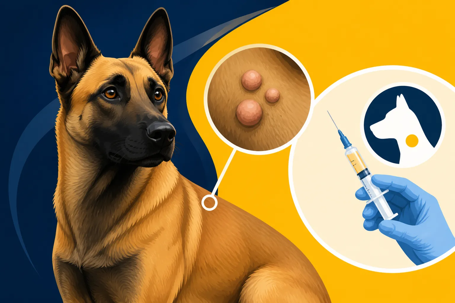
Lumps Worth Aspirating on a Belgian Malinois
Finding a lump on your Belgian Malinois can send any owner into a spiral of worry. These incredibly active and lean dogs often develop bumps and growths that might seem concerning, and it's a completely natural reaction to immediately fear the worst. Knowing which ones truly warrant immediate veterinary attention, especially the diagnostic procedure of aspiration, is absolutely key to managing your stress and your dog's health. This comprehensive guide will walk you through understanding different types of lumps, when aspiration is recommended, what the procedure entails, how to interpret the results, and what to do next.
The Immediate Worry of Finding a Lump on Your Malinois
That moment you run your hand over your Malinois’s sleek, short coat and feel an unexpected bump can stop your heart. For owners of such high-drive, athletic dogs, the first thought often jumps to injury or something far more sinister. It's a valid concern, given their intense physicality and the potential for various health issues.
Why Malinois Owners Feel Extra Concern
Belgian Malinois are not like many other breeds where a bit of extra fluff might hide a small growth for a while. Their incredibly lean and muscular physique means there’s very little fat to pad things, so even a small lump can feel quite prominent and alarming. Their active lifestyles also mean they're prone to bumps and scrapes, making it hard to discern a benign injury from something more serious. We put these dogs through so much, whether in sport, work, or just daily life, and any physical change immediately raises questions about their well-being and continued ability to perform. The breed is generally robust, but like all dogs, they are not immune to growths, and the thought of cancer is always lurking in the back of an owner's mind, especially when considering The Cancers That Take a Belgian Malinois.
Common Areas Where Lumps Appear
Lumps can pop up almost anywhere on your Malinois, but some areas are more common than others. You might find them:
- On the Trunk and Limbs: These are frequently sites for fatty tumors (lipomas) or cysts, but also where injection site reactions can occur.
- Around Joints and Pressure Points: Areas like elbows, hocks, or hips might develop hygromas (fluid-filled sacs from chronic trauma) or calluses, which can sometimes be confused with solid growths.
- On the Head and Neck: Sebaceous cysts, warts, or even swollen lymph nodes can appear here.
- Under the Skin, but Attached: Some masses, like mast cell tumors, can feel mobile under the skin but are actually adhered to deeper tissues.
- Within Muscles or Deeper Tissues: These are often harder to detect until they become quite large, and might indicate sarcomas or other muscular growths.
Given their short coat, these lumps are often visible, or at least easily felt, which is a double-edged sword: it allows for early detection but can also heighten anxiety.
Differentiating Benign from Potentially Malignant Lumps
Not all lumps are created equal, and understanding the general categories can help you communicate effectively with your vet, even if you’re not making a diagnosis yourself. The vast majority of lumps found on dogs are benign, but distinguishing them from malignant ones is a job for diagnostics, primarily fine needle aspiration or biopsy.
Understanding Lipomas and Cysts
These are two of the most common types of benign lumps you'll encounter on a Malinois, or any dog for that matter.
- Lipomas: These are simply fatty tumors. They’re usually soft, movable, and non-painful. You might be able to shift them around under the skin. They grow slowly and often appear in middle-aged to older dogs. While generally harmless, very large lipomas can sometimes interfere with movement or become unsightly. They’re typically not dangerous, but they can sometimes be tricky to distinguish from other soft tissue masses without aspiration. Often, if a lipoma is confirmed and isn’t bothering the dog, it’s simply monitored.
- Sebaceous Cysts: These occur when a sebaceous gland (which produces skin oils) becomes blocked. They often feel firm, are typically well-defined, and can be anywhere from pea-sized to golf ball-sized. They might feel a bit like a pimple under the skin, and sometimes, if ruptured, a white, pasty, or cottage cheese-like substance will come out. While benign, they can become infected or irritated, especially if your Malinois is constantly rubbing or scratching at them. These are generally more of a nuisance than a severe health threat, but infection can be painful.
When to Suspect Something More Serious
While many lumps are benign, some signs should immediately raise a red flag and prompt a visit to the vet for further investigation, such as Lumps Worth Aspirating on a Belgian Malinois.
- Rapid Growth: Any lump that seems to pop up out of nowhere or grows noticeably larger in a short period (days to weeks) needs urgent attention.
- Firmness and Fixation: If a lump feels hard, irregular, or is firmly attached to underlying tissues (muscle, bone) rather than moving freely under the skin, it’s more concerning.
- Ulceration or Bleeding: Lumps that are open, raw, or bleeding are always worrying, as this can indicate aggressive growth or infection.
- Pain or Discomfort: If your Malinois flinches, limps, or otherwise shows discomfort when the lump is touched, or if it impacts their mobility or behavior, it's a definite cause for concern.
- Location: While not a definitive indicator, certain locations, such as near lymph nodes, in the mouth, or on internal organs (like Splenic Tumors in an Older Belgian Malinois), are always more serious.
Mast cell tumors are a prime example of a lump that can vary wildly in appearance but often warrants serious attention. They can look like anything from a benign wart to a rapidly growing, ulcerated mass. This is why any new or changing lump, especially on a Malinois, should be evaluated by a professional.
The Importance of Early Detection
This isn’t just a saying; it’s a critical reality, particularly with a breed as active and predisposed to certain conditions as the Malinois. The earlier a potentially malignant lump is detected, the better the prognosis and the wider the range of treatment options available. A small, early-stage tumor is often much easier to remove completely with clear margins than a large, invasive one. Early detection can mean the difference between a simple surgical removal and a more complex, costly, and potentially less successful treatment regimen. Regularly performing "body checks" on your Malinois – feeling over their entire body, paying attention to any new bumps or changes – is one of the most proactive steps you can take for their long-term health. Don’t wait until a lump is bothering your dog or has grown significantly; if you find something, get it checked out sooner rather than later.
Deciding When to Aspirate: Key Indicators
So, you’ve found a lump. Now what? Your vet will assess the lump and, in many cases, will recommend a fine needle aspiration (FNA). This diagnostic tool is often the first step in understanding what you’re dealing with, but the decision to aspirate isn’t always cut and dry.
Size, Location, and Growth Rate
These three factors are usually the primary indicators for whether your vet will suggest aspiration.
- Growth Rate: This is arguably the most critical factor. A lump that has appeared suddenly and is growing quickly is far more suspicious than one that has been present for months or years with no change. If you notice a lump that wasn’t there last week, or that has doubled in size in a month, get it checked immediately. Even a seemingly benign cyst can become problematic if it's growing aggressively or is prone to rupture.
- Size: While size alone doesn't always denote malignancy, larger lumps can be more challenging to remove surgically if they turn out to be cancerous. Aspirating a smaller lump can give you vital information that might guide a less invasive or more successful treatment plan.
- Location: Lumps in certain areas are inherently more concerning. A small lump on a paw pad, for instance, might interfere with walking and become ulcerated, even if benign. A lump near a joint could restrict movement. And, as mentioned, growths around lymph nodes or in sensitive areas of the body always warrant immediate investigation. If your Malinois has a lump in a place where it might be easily injured during training, like a Broken Teeth on a Malinois From Bite Work or a pad injury, it might also become irritated and mimic a more serious lesion.
How to Monitor Lumps at Home
While your vet is the expert, you play a crucial role in monitoring any lumps your Malinois develops. This information can be incredibly helpful for your vet in making diagnostic decisions.
- Measure Regularly: Use a flexible tape measure or calipers to measure the length, width, and height of the lump. Record these measurements weekly, along with the date.
- Take Photos: Take clear, well-lit photos of the lump from the same angles each time. This provides a visual record of any changes in size, shape, color, or texture. Place a coin or ruler next to the lump for scale.
- Note Changes in Texture and Mobility: Is it getting harder or softer? More or less mobile under the skin? Is it becoming attached to underlying tissues?
- Observe Behavior: Is your Malinois licking, chewing, or scratching at the lump? Is it causing them pain or discomfort? Is it affecting their ability to move, eat, or play?
- Keep a Journal: Document all your observations, measurements, and photos. This detailed history is invaluable for your veterinarian.
This meticulous home monitoring can often help determine the urgency of aspiration and provide crucial context for the vet.
Your Vet's Role in Initial Assessment
When you bring your Malinois in, your veterinarian will perform a thorough physical examination, paying close attention to the lump. They'll palpate it, assessing its size, shape, consistency (soft, firm, hard), mobility (is it fixed or does it move freely?), and any tenderness or pain. They'll also check regional lymph nodes and listen to your Malinois’s overall health.
Based on this initial assessment, combined with your history of the lump (how long it’s been there, how quickly it grew, any changes you've observed), your vet will recommend the most appropriate next step. In most cases, if there's any uncertainty about the nature of the lump, a fine needle aspiration is recommended as a relatively non-invasive first diagnostic step. They might also suggest other diagnostics, like bloodwork (to check overall health or for markers of inflammation/infection) or even imaging, especially for lumps that are deep or in complex areas. If you have pet insurance like Healthy Paws, it’s a good idea to check your coverage for these diagnostic steps, as they can help alleviate financial stress during an already anxious time.
The Fine Needle Aspiration (FNA) Procedure in Detail
The thought of needles can be daunting for both you and your Malinois, but a fine needle aspiration (FNA) is a quick, minimally invasive, and generally well-tolerated procedure that provides invaluable information about lumps.
Preparing Your Malinois for Aspiration
In most cases, very little preparation is needed for an FNA. It's often done during a regular vet appointment.
- No Fasting Usually Required: Unless your vet anticipates needing sedation for an anxious or painful dog, there's typically no need to withhold food or water.
- Managing Anxiety: For highly energetic or sensitive Malinois, your vet might suggest a mild sedative to help them relax during the procedure. This is more about ensuring the dog stays still so the vet can get a good sample, rather than managing pain, as the procedure itself is quick. If your Malinois is particularly reactive to new environments or being handled, discuss this with your vet beforehand. Sometimes, simple counter-conditioning with high-value treats can make a world of difference, especially for a food-motivated Malinois.
- Cleanliness: The area around the lump might be gently cleaned with an antiseptic solution, but often hair isn’t even shaved for a simple FNA.
The goal is to make the experience as stress-free as possible for your Malinois. Remember, these dogs are smart and can pick up on your anxiety, so staying calm yourself is helpful.
What Happens During the Aspiration
A fine needle aspiration is exactly what it sounds like: using a very thin needle to collect a sample of cells from the lump.
- Immobilization: Your Malinois will be gently restrained by a vet tech or assistant. For most Malinois, a firm but reassuring hold is enough.
- Needle Insertion: Your vet will carefully insert a small, hollow needle (similar to one used for vaccinations) directly into the lump. They may move the needle around within the mass a few times to collect a representative sample of cells. This "fanning" technique ensures they get cells from different areas of the lump.
- Aspiration: Often, a syringe attached to the needle is used to create a vacuum, gently suctioning cells into the needle hub. Sometimes, just the needle itself is enough to collect cells through capillary action.
- Sample Collection: The needle is then withdrawn. The collected material, which is often just a tiny speck, is gently expelled onto a microscope slide. Your vet will then spread the cells thinly across the slide, creating a "smear."
- Multiple Samples: Often, several aspirations are performed from different areas of the lump to increase the chances of getting a diagnostic sample. This is particularly important for heterogeneous masses, such as Mast Cell Lumps Found on a Working Malinois, which can have varying cell types within them.
The entire process is usually over in a minute or two. Your Malinois might feel a brief prick, similar to a vaccine, and perhaps a strange pressure sensation as the needle moves within the lump. Most dogs tolerate it remarkably well.
Is it Painful? Addressing Owner Concerns
This is a common and understandable question for any dog owner. For the vast majority of Malinois, an FNA causes only minimal discomfort, akin to a quick pinprick.
- Minimal Pain: The needles used are very fine, and the procedure is quick. Most dogs react with a slight flinch or don't even seem to notice it.
- Local Anesthetic Rarely Needed: While a local anesthetic could be used, it's rarely necessary for a simple FNA because the injection of the anesthetic itself can be as uncomfortable as the aspiration, and it adds another step to the procedure.
- Post-Procedure: Your Malinois might have a tiny bit of bruising or mild soreness at the site, but this usually resolves very quickly, often within hours. There’s typically no need for pain medication afterwards.
If your Malinois is particularly sensitive or the lump is in a very tender area, your vet might discuss options for light sedation. However, for most Malinois, their curiosity or high drive often distracts them enough that they don’t fuss much. The biggest risk isn’t pain, but rather the dog moving unexpectedly and making it harder for the vet to get a good sample.
What the Aspiration Results Can Tell You
Once the samples are collected, they are prepared and sent to a veterinary pathologist, a specialist trained in interpreting cell samples. The results of the aspiration can provide crucial insights, guiding the next steps in your Malinois's care.
Interpreting the Cytology Report
The pathologist examines the cells under a microscope and compiles a cytology report, which your vet will then discuss with you. This report typically categorizes the cells found, helping to identify the type of lump.
Common findings include:
- Fat Cells: This is the most common finding for a lipoma. These cells are distinctive and easily identifiable, confirming the lump is a benign fatty tumor.
- Inflammatory Cells: The presence of neutrophils, macrophages, or other immune cells might indicate an abscess (infection), a foreign body reaction (e.g., a splinter), or an inflammatory cyst. If an abscess is identified, the vet might drain it or prescribe antibiotics.
- Epithelial Cells: These are skin cells. Depending on their appearance and arrangement, they could indicate a benign cyst, a wart, or in some cases, a carcinoma (a type of malignant tumor).
- Mesenchymal Cells (Spindle Cells): These cells can be found in a variety of soft tissue masses, both benign and malignant (e.g., fibromas, sarcomas). Their appearance and arrangement help the pathologist determine if they are concerning.
- Mast Cells: The presence of mast cells is significant as it indicates a mast cell tumor, which can be highly variable in its malignancy but always requires attention. Depending on the grade of the cells, further action like surgical removal and potentially chemotherapy might be recommended. This is a common and important finding that dictates immediate action, and you'll want to address it, especially if thinking about The Bleeding Tumor That Kills an Older Malinois.
- Melanocytes: Pigment-producing cells. While many melanomas are benign, some can be aggressive, particularly in certain locations like the mouth or nail beds.
The report will often provide a diagnosis or at least narrow down the possibilities, helping your vet formulate a plan.
When Aspiration is Inconclusive
It's important to understand that while FNA is a powerful tool, it does have its limitations. Sometimes, the results can come back as "inconclusive" or "non-diagnostic." This can be frustrating, but it doesn't mean the procedure was a waste; it simply means more information is needed.
Reasons for an inconclusive result can include:
- Insufficient Sample: Sometimes, not enough cells are collected, or the cells rupture during collection, making them unreadable.
- Blood Contamination: Too much blood in the sample can obscure the cells.
- Firm or Scarred Tissue: Very firm or fibrous lumps, like some sarcomas, don't exfoliate cells well, meaning the needle can't pick up enough diagnostic material.
- Necrotic Tissue: If the center of a tumor is dying, the collected cells may not be representative of the main mass.
- Heterogeneous Masses: Some tumors have different types of cells throughout, and the sample might only capture one section, missing the malignant cells.
If an aspiration is inconclusive, your vet will discuss alternative diagnostic steps. This most often means a biopsy, which involves surgically removing a larger piece of the lump (incisional biopsy) or the entire lump (excisional biopsy) for histopathology. Histopathology provides a tissue architecture view, which is far more comprehensive than just a cellular view, and is considered the gold standard for diagnosing many tumors.
Next Steps Based on Diagnosis
The path forward after aspiration depends entirely on the cytology report:
- Benign (e.g., Lipoma, simple cyst): Your vet might recommend simply monitoring the lump for any changes, especially if it's not bothering your Malinois. For an infected cyst or a large lipoma causing issues, surgical removal might still be an option.
- Inflammatory/Infectious (e.g., Abscess): Treatment might involve antibiotics, anti-inflammatory medications, draining the abscess, or removing a foreign body.
- Malignant (e.g., Mast Cell Tumor, Carcinoma, Sarcoma): This is where quick action is often vital. The primary recommendation is usually surgical removal with wide margins to ensure all cancerous cells are excised. Depending on the type and grade of cancer, further diagnostics (like imaging to check for spread) or treatments (such as chemotherapy or radiation) might be advised. Discussions about When a Lump on a Malinois Needs Removing will definitely be at the forefront of your conversations with your vet.
- Inconclusive: As discussed, a biopsy is typically the next logical step to get a definitive diagnosis.
Your vet will guide you through these options, discussing the pros, cons, costs, and prognosis for each. This is where having pet insurance like Healthy Paws can truly be a lifesaver, covering a significant portion of diagnostic and treatment costs.
Living With Lumps: Management and Prevention
Even after a lump is diagnosed, the journey often isn't over. Whether it's ongoing monitoring, post-surgical care, or simply a proactive approach to health, there are crucial steps to take as a responsible Malinois owner.
Post-Aspiration Care and Monitoring at Home
After your Malinois has had an FNA, the immediate aftercare is usually minimal, but continued vigilance is important.
- Keep the Site Clean: The tiny puncture wound typically heals very quickly. You might be advised to gently clean the area with a mild antiseptic wipe, but usually, just keeping an eye on it is sufficient. Prevent your Malinois from licking or chewing the site, especially if there's any tenderness. An Elizabethan collar (cone) might be necessary for a short period if they're persistent.
- Watch for Changes: Even if a lump was diagnosed as benign, it's wise to continue your home monitoring routine. Check the lump regularly for any changes in size, shape, color, or texture. Are there new lumps appearing? Are existing ones growing or changing? This continued observation is critical because a benign lump can sometimes still grow to a problematic size, or a new, more serious lump could appear elsewhere.
- Follow-Up Appointments: Adhere to any follow-up schedule your vet recommends. This might be a recheck in a few weeks or months, or an immediate follow-up if further diagnostics or treatment are planned.
Remember, your Malinois relies on you to be their advocate. Your observations and diligence are invaluable in their health management.
Proactive Health Measures for Malinois
Beyond just reacting to lumps, there are broader health practices that can support your Malinois's overall well-being and potentially reduce the incidence or severity of certain conditions.
- Regular Veterinary Check-ups: This cannot be stressed enough. A thorough annual (or bi-annual for older dogs) exam allows your vet to catch subtle changes you might miss. They can perform a full body palpation, check lymph nodes, and discuss any new findings. These routine visits are part of The Checkup Schedule a Sport Malinois Should Keep and are critical for early disease detection.
- Balanced Nutrition: A high-quality diet appropriate for an active Malinois supports a strong immune system and healthy cellular function. Discuss with your vet if any supplements might be beneficial for your dog's specific needs, though generally, a balanced commercial diet is sufficient.
- Weight Management: Keeping your Malinois at a lean, healthy weight is vital. Excess weight can exacerbate joint issues, contribute to other health problems, and potentially make lumps harder to detect.
- Environmental Awareness: Be mindful of chemicals in your home or yard, as some environmental toxins have been linked to certain cancers in dogs. While it's hard to eliminate all risks, minimizing exposure can be beneficial.
- Recognizing Behavioral Changes: A Malinois is a creature of habit. Any subtle shifts in energy levels, appetite, thirst, bathroom habits, or willingness to work could be an early sign of an underlying health issue, not necessarily just a lump.
By combining proactive general health management with targeted vigilance for lumps, you’re providing the best possible care for your dedicated companion.
The Role of Pet Insurance in Managing Lumps
Let’s be realistic: vet bills, especially for diagnostics and treatment of potentially serious lumps, can add up quickly. This is where pet insurance, like a plan from Healthy Paws, truly shines.
- Financial Peace of Mind: Knowing that a significant portion of diagnostic tests (like FNA, biopsies, imaging), surgeries, medications, and even specialized treatments (like chemotherapy) can be covered by your insurance policy reduces the immense financial stress that often accompanies a cancer diagnosis or the need for lump removal.
- Access to Best Care: Without the burden of upfront costs, you're free to make decisions based on what’s best for your Malinois’s health, rather than your wallet. This means you’re more likely to pursue every recommended diagnostic and treatment option, increasing your dog's chances of a positive outcome. Consider What Pet Insurance Runs on a Working Malinois to understand the investment, but the payoff can be invaluable.
- Coverage for the Unexpected: While we hope our Malinois never face a serious health crisis, lumps and bumps are a common occurrence in dogs. Having insurance ensures you’re prepared for these unexpected events, whether it’s a simple infected cyst or something far more complex.
It's always a good idea to enroll your Malinois in pet insurance when they are young and healthy, as pre-existing conditions are typically not covered. This ensures that when a lump does appear, whether at age two or ten, you're covered for the necessary steps to diagnose and treat it, ultimately giving your Malinois the best chance at a long, healthy life.
Navigating Malinois Lumps: A Summary of Common Types and Actions
To help organize your thoughts when that worrying lump appears, here's a quick reference guide on some common types of lumps and the general approach:
| Lump Type | Common Characteristics | Typical Feel | Growth Rate | Aspiration Often Suggested | Next Steps (Post-Aspiration) |
|---|---|---|---|---|---|
| Lipoma | Fatty tumor, usually under skin, non-painful | Soft, movable | Slow | Often elective, for confirmation | Monitor, or surgical removal if large/interfering. |
| Sebaceous Cyst | Blocked oil gland, can rupture pasty material | Firm, well-defined | Slow | If ruptured, infected, or for confirmation. | Monitor, treat infection, or surgical removal if problematic. |
| Histiocytoma | Benign skin tumor, common in young dogs, often on head/limbs | Firm, dome-shaped | Rapid, then often regresses | Often for confirmation, or to rule out mast cell tumor. | Monitor (often regresses on its own), or surgical removal. |
| Abscess | Collection of pus, often from infection/puncture wound | Soft/firm, warm, painful, can be fluctuant | Rapid | Essential, for diagnosis and culture to guide antibiotics. | Drain, antibiotics. |
| Mast Cell Tumor | Highly variable, can look like anything from wart to ulcerated mass | Variable, often firm, can be fixed or mobile | Variable, can be rapid | Essential, critical for early diagnosis and grading. | Surgical removal with wide margins, further diagnostics, potential chemotherapy. |
| Injection Site Reaction/Granuloma | Localized swelling after vaccine/medication | Firm, sometimes painful | Variable | For confirmation, to rule out other issues. | Monitor, anti-inflammatories. |
This table serves as a general guide, but always consult your veterinarian for any new or changing lump on your Belgian Malinois.
Disclaimer
The information provided in this article is for general informational purposes only and is not a substitute for professional veterinary advice, diagnosis, or treatment. Always seek the advice of your veterinarian or other qualified animal health provider with any questions you may have regarding a medical condition or treatment for your Belgian Malinois.