When a Lump on a Malinois Needs Removing
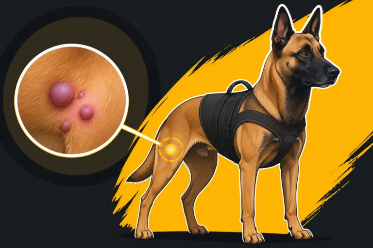
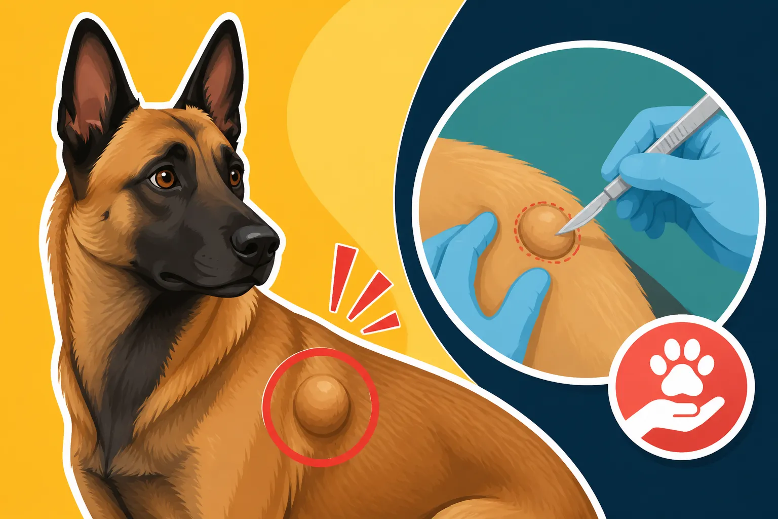
Finding an unexpected lump on your Belgian Malinois can send a bolt of fear straight through any dedicated owner. One moment you're petting their sleek, muscled form, and the next your fingers brush against something that absolutely shouldn't be there. It's a terrifying moment, and the immediate worry is always "What if it's cancer?" This guide is here to walk you through the practical steps and considerations for when a lump on your Malinois needs to be evaluated and potentially removed, helping you navigate that anxious period with a clear head.
The Initial Discovery: Panic, Poking, and Practical First Steps
That sinking feeling when you discover an unusual bump on your Malinois is completely normal. Our dogs are family, often our working partners, and any deviation from their robust health immediately sparks concern. It's easy to fall into a rabbit hole of online searches or panic, but a systematic approach will serve both you and your Malinois best.
The Immediate Fear and Why It's Normal
Let's be real, a Malinois isn't just any dog. They're usually high-drive, lean machines, and any soft spot or odd protrusion feels inherently wrong on their athletic build. When you feel that lump, your mind probably jumps to the worst-case scenario. Is it a tumor? Is it something aggressive that's been growing undetected? This immediate fear is a natural response to the unknown, especially given how much we invest in these incredible dogs. You’re not alone in feeling this way. Every Malinois owner who has found a lump has likely felt that same gut-punch of anxiety. It's tough to keep a level head, but remembering that not all lumps are sinister can offer a small comfort as you move forward.
What to Note Down When You Find a Lump
Before you even pick up the phone to call the vet, take a moment to observe and document. This isn't just for your own peace of mind; it provides crucial information for your veterinarian. Think of yourself as a detective. What did you notice?
- Location: Be precise. Is it on their leg, chest, flank, or near an ear? Is it on the ribs, or further back? On a sensitive area like the groin? Knowing the exact spot helps the vet during examination and for future monitoring.
- Size: How big is it? Compare it to something common, like a pea, a marble, a grape, or a golf ball.
- Texture: Is it hard, soft, squishy, firm? Does it move freely under the skin, or does it feel attached to underlying tissue?
- Appearance: Is the skin over it normal, red, inflamed, or losing hair? Is there any discharge?
- Growth: When did you first notice it? Has it changed in size or appearance since then? Has it grown rapidly, or slowly?
- Pain/Discomfort: Does your Malinois react when you touch it? Do they seem bothered by it in their daily activities?
- Other Symptoms: Has your Malinois been acting differently? Lethargic, loss of appetite, changes in energy or working drive? Even subtle changes can be relevant.
This detailed information will streamline your vet visit and give them a much better starting point.
When to Schedule That Vet Visit
Honestly, the moment you find a new, unexplained lump on your Belgian Malinois, it's time to call the vet. While it might turn out to be nothing serious, prompt evaluation is always the safest course of action. Rapidly growing lumps, or those that appear inflamed, painful, or accompanied by other signs of illness, warrant an urgent visit, ideally within a day or two. If the lump is small, seems stable, and your Malinois is otherwise acting completely normal, a regular appointment within the week is generally acceptable. However, do not delay. Even seemingly innocent lumps can sometimes mask something more serious, and catching potential issues early can make a world of difference in prognosis and treatment options. Think of it as part of their routine care, much like ensuring they have The Checkup Schedule a Sport Malinois Should Keep.
Common Causes of Lumps on a Belgian Malinois
It’s easy for our minds to jump straight to the worst-case scenario when we find a lump. However, not every lump on your Belgian Malinois is a death sentence. There's a wide spectrum of possibilities, ranging from completely harmless to very serious, and understanding these can help temper your anxiety while awaiting a diagnosis.
Benign Bumps: The "Just a Thing" Lumps
Many lumps found on dogs, including active breeds like the Belgian Malinois, turn out to be benign. These are growths that aren't cancerous and don't spread to other parts of the body.
- Lipomas: These are fatty tumors and are incredibly common, especially in older dogs. They feel soft and mobile under the skin. While they can sometimes grow quite large and interfere with movement or be unsightly, they are generally harmless. You might find one on their chest, flank, or even their belly. They usually don't bother the dog unless they're in a high-friction area or become excessively large.
- Cysts: Cysts are sacs filled with fluid, dead cells, or other material. They can be follicular cysts, sebaceous cysts (blocked oil glands), or even apocrine cysts. They often feel somewhat soft and can sometimes rupture, leading to irritation or infection. They might appear anywhere, including on the head or neck.
- Warts (Papillomas): These are usually viral and look like small, cauliflower-like growths. They are more common in younger dogs but can appear at any age. While generally harmless, some can be irritating if located where they rub.
- Histiocytomas: These are benign skin tumors most often seen in young dogs, particularly on the head, ears, or limbs. They tend to grow rapidly but often regress and disappear on their own within a few months. Your vet can usually diagnose these based on their appearance and location, though an FNA can confirm.
These "just a thing" lumps might need monitoring or minor intervention if they're causing issues, but they don't carry the grave prognosis of malignant growths.
Inflammatory Reactions and Infections
Sometimes, a lump isn't a growth at all, but rather a reaction from the body to an irritant, injury, or infection.
- Abscesses: These are collections of pus under the skin, often caused by a bite wound, puncture, or other skin trauma that introduces bacteria. They can be hot to the touch, painful, and may cause fever or lethargy. They often feel firm and then become softer as the pus accumulates, eventually rupturing if not drained. Given a Malinois's active lifestyle, running through brush or engaging in bite work, minor injuries that can lead to abscesses aren't uncommon.
- Granulomas: These are masses of immune cells that form in response to chronic inflammation or infection. They can be sterile (non-infectious) or infectious, often in response to foreign bodies (like a splinter or a foxtail), fungal infections, or even some parasitic infestations. They can appear anywhere on the body and vary in size and firmness.
- Insect Bites or Stings: A bee sting or an insect bite can cause a localized swelling that feels like a lump, often accompanied by redness and tenderness. These usually resolve on their own, but some dogs can have more significant allergic reactions.
- Reactions to Injections: Occasionally, a small, firm lump can form at the site of a recent injection (vaccine, medication). These are typically benign and resolve over time, but it's always good to point them out to your vet, especially if they persist. For Malinois that might be sensitive, like those where you need to consider Whether a Malinois Reacts to Vaccines, this can be a particular point of awareness.
These types of lumps often require treatment for the underlying cause, such as antibiotics for an abscess or anti-inflammatories for a reaction, rather than surgical removal of the lump itself.
The C Word: When Lumps Are More Serious
Unfortunately, some lumps do turn out to be malignant (cancerous). Catching these early is paramount for a better outcome.
- Mast Cell Tumors (MCTs): These are one of the most common skin tumors in dogs and can appear anywhere on the body, including internal organs. They are highly variable in appearance – they can be firm, soft, raised, flat, red, or hairless. They can also change in size, sometimes fluctuating dramatically (a phenomenon known as Darier's sign). Mast cells release histamine, which can cause local irritation, redness, and swelling. Due to their unpredictable nature and potential for spread, prompt diagnosis and aggressive treatment are crucial.
- Soft Tissue Sarcomas (STSs): These are a group of malignant tumors that arise from connective tissues. They typically feel firm and are often fixed to underlying tissues, making them less mobile than a lipoma. They can occur anywhere but are common on limbs and the trunk. Wide surgical margins are often needed for successful removal.
- Melanomas: While often benign in dogs, malignant melanomas can be aggressive, particularly when found in certain locations like the mouth, nail beds, or mucocutaneous junctions (where skin meets mucous membrane). They are usually dark pigmented but can sometimes be amelanotic (lacking pigment).
- Lymphoma: This cancer of the lymphatic system can present as enlarged lymph nodes, which feel like firm, non-painful lumps, particularly in the neck (submandibular), armpits (axillary), or groin (inguinal). However, it can also manifest as skin lesions.
- Carcinomas: These cancers arise from epithelial cells and can occur in various forms, such as squamous cell carcinoma or basal cell carcinoma. Their appearance can vary widely, from ulcerated lesions to firm nodules.
Given the potential severity of these, it’s clear why finding a lump immediately prompts concern. This is why a thorough diagnostic process, often starting with procedures mentioned in articles like Lumps Worth Aspirating on a Belgian Malinois, is so critical.
Diagnostic Steps: Beyond the Visual Exam
While your vet can learn a lot from simply looking at and feeling a lump, a definitive diagnosis almost always requires further investigation. This is where medical procedures come into play, providing the crucial information needed to decide on the best course of action.
Fine Needle Aspirate FNA The First Line of Defense
The Fine Needle Aspirate (FNA) is often the first, least invasive, and most cost-effective diagnostic step. It involves using a small needle, similar to one used for vaccinations, to collect a sample of cells from the lump. The collected cells are then spread onto a glass slide and examined under a microscope by a veterinarian or a veterinary pathologist.
- How it works: Your vet will gently insert the needle into the lump, move it back and forth a few times to gather cells, and then withdraw it. It's usually a quick procedure that many Malinois tolerate without sedation, though a super high-strung dog might need a quick muzzle or a second person to help hold them steady.
- What it tells you: An FNA can often distinguish between fatty tumors (lipomas), cysts, inflammatory cells (like those seen in abscesses or granulomas), and some types of cancerous cells (like mast cells). It helps your vet get a preliminary idea of what they're dealing with.
- Limitations: It's important to understand that an FNA isn't always definitive. Sometimes, the sample collected might not be representative of the entire lump (if it's heterogeneous), or the cells might be too few or too damaged to yield a clear diagnosis. In such cases, the result might come back as "non-diagnostic," meaning more advanced testing is needed. It’s a great screening tool but not always the final word.
Biopsy and Histopathology: Getting a Definitive Answer
If an FNA is inconclusive, or if the vet suspects a more serious lump based on its appearance or behavior, a biopsy is the next step. A biopsy involves surgically removing a larger piece of tissue, or the entire lump, which is then sent to a specialized veterinary pathologist for histopathology – a detailed examination of the tissue structure.
- Types of biopsies:
- Incisional Biopsy: Only a portion of the lump is removed. This is often done for very large lumps or those in tricky locations, where removing the entire thing initially might be too invasive without a definitive diagnosis.
- Excisional Biopsy: The entire lump, along with a margin of healthy tissue around it, is removed. This is often preferred if the lump is small and easily accessible, as it can be both diagnostic and curative in one go.
- What it tells you: Histopathology provides the most accurate diagnosis. The pathologist can identify the specific type of cells, determine if the growth is benign or malignant, grade the tumor (how aggressive it appears), and assess whether surgical margins are clear (meaning the entire tumor was removed). This level of detail is critical for developing an appropriate treatment plan.
- Anesthesia: A biopsy, even an incisional one, requires local anesthetic at minimum, but often general anesthesia, especially if the Malinois is squirmy, the lump is deep, or a larger sample is needed. Given the athletic and often high-strung nature of a Malinois, vets are always careful with Anesthesia Protocols for a Lean Belgian Malinois, making sure to discuss everything like What to Tell a Vet Before a Malinois Is Sedated.
Additional Imaging and Staging
For malignant lumps, especially those with a high potential for spread, your vet might recommend further imaging and staging to determine if the cancer has spread to other parts of the body.
- Radiographs (X-rays): Chest X-rays are commonly performed to check for metastasis (spread) to the lungs, a common site for many canine cancers. Abdominal X-rays might be taken if internal organ involvement is suspected.
- Ultrasound: An abdominal ultrasound can be used to examine internal organs like the spleen, liver, and lymph nodes for signs of metastasis. This is particularly relevant if the lump is a type known to spread internally, such as a splenic tumor that might lead to The Bleeding Tumor That Kills an Older Malinois.
- CT Scan or MRI: In more complex cases, especially with lumps in difficult anatomical locations or those suspected to have invaded deeper tissues, advanced imaging like CT scans or MRIs might be recommended. These provide highly detailed cross-sectional images, helping surgeons plan complex removals.
- Bloodwork: While not directly diagnosing the lump, pre-anesthetic bloodwork is essential before any surgical procedure, and broader blood panels can sometimes reveal systemic issues or give clues about a dog's overall health, particularly if they’ve had The Bloodwork a Working Malinois Should Have Annually.
All these steps contribute to painting a full picture, allowing you and your vet to make the most informed decision possible for your Malinois.
Deciding on Removal: Factors to Consider
Once you have a diagnosis, the big question becomes: should this lump be removed? This decision isn't always straightforward and involves weighing several factors, including the nature of the lump, your dog's overall health, and practical considerations.
Location, Size, and Growth Rate
The characteristics of the lump itself play a huge role in the decision-making process.
- Location: A lump on the side of the chest might be easier to remove than one on a joint, near a major nerve, or in an area with minimal extra skin, like the lower leg. Lumps in high-motion areas or those that rub (e.g., in the armpit or groin) might cause discomfort and irritation, making removal more appealing even if benign. Lumps around the face or mouth can quickly become problematic for eating, breathing, or vision.
- Size: Smaller lumps are generally easier to remove with clean margins and less tissue disruption. If a lump is benign but growing very large, it might eventually interfere with movement or quality of life, making removal a good preventative measure. For malignant lumps, smaller size often means less likelihood of spread and a better surgical outcome.
- Growth Rate: A lump that is rapidly growing is always more concerning and often warrants prompt removal and aggressive diagnosis. Even if an FNA suggests it's benign, rapid growth can sometimes indicate a more aggressive subtype or a misdiagnosis, making excision the safest bet. A stable, slow-growing lump, especially if diagnosed as a benign lipoma, might simply be monitored.
The Malinois's Age, Health, and Temperament
Your Malinois isn't just a lump; they're an individual with their own set of health considerations.
- Age: An older Malinois, while still likely robust, might have other health issues that make anesthesia and surgery riskier. For a very elderly dog with multiple comorbidities, a benign lump that isn't bothering them might be left alone, whereas a younger dog would likely have it removed. Conversely, certain aggressive cancers can be particularly devastating in younger dogs, making prompt and decisive action more critical.
- Overall Health: Any pre-existing conditions, such as heart murmurs, kidney disease, or a history of Managing an Epileptic Belgian Malinois on Medication, will factor into the risk assessment for anesthesia and surgery. Your vet will conduct a thorough pre-surgical workup to identify any potential complications.
- Temperament: A Malinois's intense drive and high-energy personality can sometimes make recovery from surgery challenging. Strict confinement and activity restriction might be incredibly difficult for a dog used to constant work and engagement. This needs to be considered, as a dog who constantly stresses and damages their incision could complicate recovery. Your vet might suggest specific pain management or mild sedatives to help during the recovery period.
Financial Considerations and Insurance
Let's be honest, vet bills can add up, and surgery for a lump can be a significant expense.
- Cost of Diagnosis and Surgery: The cost varies widely depending on the lump's complexity, the need for advanced imaging, the type of biopsy, the skill of the surgeon, and geographic location. Basic FNAs might be a couple of hundred dollars, but a full excisional biopsy with histopathology and staging could easily run into thousands. Factors like the difficulty of the surgery (e.g., removing a lump near a crucial nerve), or the need for a specialist, will drive up costs significantly. Just like how The Cost of Repairing a Malinois's Broken Canine can be quite high, any major surgical procedure on a Malinois can lead to substantial expenses.
- Pet Insurance: This is where pet insurance can be a lifesaver. Companies like Healthy Paws can help offset the financial burden of unexpected veterinary care, including diagnostics and surgery for lumps. If you have a plan with good coverage, it can certainly make the decision to pursue removal much easier, as it alleviates some of the stress of high out-of-pocket costs. Many policies, including those from Healthy Paws, cover eligible accidents and illnesses, which would include the investigation and treatment of suspicious lumps.
- Payment Plans and Grants: If you don't have insurance or face high out-of-pocket costs, discuss payment options with your veterinary clinic. Many offer payment plans, or you might look into veterinary credit options like CareCredit. There are also various charitable organizations and grant programs designed to assist owners with veterinary expenses, particularly for life-saving treatments.
This table provides a summary of key factors to consider:
| Factor | Benign Lump (e.g., Lipoma, Small Cyst) | Malignant Lump (e.g., Mast Cell Tumor, Sarcoma) |
|---|---|---|
| Growth Rate | Slow or stable | Rapid, fluctuating, or unpredictable |
| Location | Interfering with movement/function, or in high-friction areas | Anywhere, but aggressive margins needed for critical areas |
| Size | Large enough to cause discomfort or become unsightly | Any size, but smaller is better for prognosis and complete removal |
| Dog's Age | Older dog: May monitor if not bothering them; younger dog: removal for aesthetics/future prevention | Any age: Removal often recommended if prognosis allows |
| Dog's Health | Consider anesthesia risks; if healthy, removal is lower risk | Consider anesthesia risks; often necessary even with some risks |
| Temperament | May need sedation for recovery if highly active | Strict recovery often crucial for healing and preventing self-trauma |
| Cost | Elective, usually less urgent | Often urgent, potentially higher costs due to complexity/staging |
| Prognosis | Excellent | Varies widely, early diagnosis and removal improve chances |
Post-Removal Care and What to Expect
Once the decision is made and the lump is removed, the focus shifts to ensuring a smooth recovery for your Malinois. This period is critical for proper healing and minimizing complications.
Managing the Incision and Activity Restrictions
The recovery period from any surgery, especially for a high-drive Malinois, requires strict management and patience.
- Incision Care: You'll typically be sent home with instructions on how to care for the incision site. This usually involves keeping it clean and dry. Avoid bathing your Malinois for at least 10-14 days, or as directed by your vet, to prevent infection and allow the incision to seal properly. You'll need to check the incision daily for any signs of redness, swelling, heat, discharge, or if the sutures appear to be coming undone. Contact your vet immediately if you notice any of these issues.
- Preventing Licking/Chewing: This is perhaps the biggest challenge with a Malinois. Their intense focus and often high pain tolerance mean they might not show obvious signs of discomfort, but they can quickly chew out sutures and reopen a wound. An Elizabethan collar (e-collar, or "cone of shame") is almost always necessary. While it can be frustrating for both you and your dog, it's non-negotiable for proper healing. There are also soft cones, recovery suits (like the Suitical), or even inflatable collars that might be more comfortable for some dogs, but ensure whatever you choose effectively prevents access to the incision.
- Activity Restriction: This is arguably the most difficult part for a Belgian Malinois owner. Your vet will likely recommend strict crate rest or very limited activity for 10-14 days, or until the sutures are removed and the incision is healed. This means no running, jumping, stairs, rough play, or off-leash exercise. Leashed potty breaks are generally the extent of their outdoor activity. For a dog that demands Two Hours and a Job a Belgian Malinois Demands Daily, this can feel like an impossible task. You might need to rely on mental stimulation, like puzzle toys, gentle training sessions, or even mild sedatives prescribed by your vet, to keep their energy in check.
- Pain Management: Your vet will prescribe pain medication to keep your Malinois comfortable during recovery. It's crucial to administer these exactly as directed. Keeping pain under control not only improves their well-being but also helps prevent them from bothering the incision site.
When to Recheck and Monitor for Recurrence
Follow-up appointments are essential to ensure everything is healing correctly and to discuss the biopsy results in detail.
- Suture Removal: Typically, sutures are removed around 10-14 days post-surgery, assuming the incision has healed well. This is a quick visit where the vet will also check the incision site thoroughly.
- Biopsy Results Discussion: If you haven't already received them, the biopsy results will be reviewed. This discussion is vital for understanding the exact nature of the lump, whether it was benign or malignant, the grade of any cancer, and whether "clean margins" were achieved (meaning all cancerous cells appear to have been removed).
- Further Treatment Options: If the lump was malignant and margins were not clear, or if the type of cancer is known to be aggressive, your vet will discuss further treatment options. This could include a second surgery to achieve wider margins, radiation therapy, chemotherapy, or other supportive care. This is a conversation where having pet insurance, such as through Healthy Paws, can greatly impact the feasibility of these often costly, but life-extending, treatments.
- Long-term Monitoring: Even for benign lumps, and especially for malignant ones, ongoing monitoring is important. You'll need to regularly check your Malinois for any new lumps, or for any signs of recurrence at the surgical site. For certain cancers, regular follow-up imaging (X-rays, ultrasounds) or bloodwork might be recommended to screen for metastasis. This commitment to continuous observation is part of being a responsible owner, especially for a breed that lives for The Fourteen to Sixteen Years a Belgian Malinois Lives and we want to ensure every year is as healthy as possible.
Navigating the journey from lump discovery to post-operative care can be stressful, but by working closely with your veterinarian and understanding each step, you can provide the best possible care for your Belgian Malinois.
Disclaimer
The information provided in this guide is for general informational purposes only and does not constitute veterinary advice. Always consult with a qualified veterinarian for any health concerns or before making any decisions about your Belgian Malinois's medical care.